
 +86-0515-86223369
  0
0

หากคุณต้องการความช่วยเหลือใด ๆ โปรดติดต่อเรา
 Products & Solutions
 {:พ.ศ}

บ้าน / ข่าว / ข่าวอุตสาหกรรม / การทำความสะอาดคิวเวตต์ควอตซ์สำหรับ CD Spectroscopy: คู่มือห้องปฏิบัติการฉบับสมบูรณ์
การทำความสะอาดคิวเวตต์ควอตซ์สำหรับ CD Spectroscopy: คู่มือห้องปฏิบัติการฉบับสมบูรณ์
สเปกตรัมไดโครอิซึมแบบวงกลมโปรตีนของคุณดูสมบูรณ์แบบเมื่อวานนี้ ปัจจุบัน เส้นฐานโค้งขึ้นไปต่ำกว่า 215 นาโนเมตร อัตราส่วนสัญญาณต่อเสียงรบกวนลดลง และโครงสร้างรองบอกว่าเกลียวอัลฟ่าลดลงจาก 40 เปอร์เซ็นต์เหลือ 25 เปอร์เซ็นต์ ไม่มีอะไรเกี่ยวกับตัวอย่างเปลี่ยนแปลง ผู้ต้องสงสัยตามปกตินั่งอยู่ในที่วางตัวอย่าง: คิวเวตต์ควอตซ์ที่ไม่สะอาด การทำความสะอาดคิวเวตต์ควอทซ์สำหรับสเปกโตรสโคปีซีดีเป็นขั้นตอนประจำที่ห้องปฏิบัติการประเมินต่ำไป และเป็นสถานที่แรกที่จะตรวจสอบเมื่อคุณภาพข้อมูลลดลงอย่างไม่คาดคิด
ข้อสรุปหลักสามารถระบุได้ตั้งแต่เริ่มแรก: คิวเวตต์ที่ดูสะอาดอย่างสมบูรณ์แบบในช่วงความยาวคลื่นที่มองเห็นได้ ยังคงสกปรกอย่างร้ายแรงที่ 190 ถึง 230 นาโนเมตร ซึ่งเป็นที่ซึ่งการวัด CD โปรตีนส่วนใหญ่เกิดขึ้น ความแตกต่างระหว่างเส้นฐานที่ใช้งานได้กับสเปกตรัมที่เสียหายมักเป็นฟิล์มบางเกินกว่าจะมองเห็นได้ คู่มือนี้จะอธิบายว่าทำไมซีดีสเปกโทรสโกปีจึงมีความไวต่อสภาวะของคิวเวตต์โดยเฉพาะ วิธีระบุสิ่งปนเปื้อนแต่ละประเภท วิธีการทำความสะอาดวิธีใดที่จะคืนสภาพพื้นผิวแสง ซึ่งข้อผิดพลาดทำให้เกิดความเสียหายถาวร และวิธีการตรวจสอบว่าคิวเวตต์พร้อมสำหรับการตรวจวัดอย่างแท้จริง
เกณฑ์วิธีด้านล่างนี้เป็นขั้นตอนปฏิบัติที่ใช้ในงานชีวฟิสิกส์โปรตีนตามปกติ สามารถใช้งานร่วมกับคิวเวตต์ควอตซ์และซิลิกาผสมจากผู้ผลิตคิวเวตต์ควอตซ์ที่มีชื่อเสียงใดๆ และได้รับคำสั่งตั้งแต่การล้างรายวันที่รุนแรงน้อยที่สุดไปจนถึงขั้นตอนการชำระล้างการปนเปื้อนที่ลึกยิ่งขึ้นซึ่งใช้เฉพาะเมื่อการทำความสะอาดตามปกติยังไม่เพียงพอ
บทสรุปที่สำคัญ: คิวเวตต์ควอตซ์ที่สะอาดเป็นวิธีประกันที่ง่ายที่สุดและถูกที่สุดสำหรับข้อมูลไดโครอิซึมแบบวงกลมที่สามารถทำซ้ำได้ ขจัดการปนเปื้อนของคิวเวตต์ทุกครั้งก่อนสัมผัสเครื่องมือ
เหตุใด CD Spectroscopy จึงต้องการความสะอาดของคิวเวทท์อย่างสูงสุด
ไดโครอิซึมแบบวงกลมวัดความแตกต่างเล็กๆ น้อยๆ ในการดูดกลืนแสงระหว่างแสงโพลาไรซ์แบบวงกลมซ้ายและขวา ย่านความถี่ CD โดยทั่วไปขนาด 10 มิลลิองศา สอดคล้องกับค่าความต่างของการดูดกลืนแสงประมาณ 3 x 10^-4 AU ซึ่งเล็กกว่าย่านการดูดกลืนแสงที่บันทึกไว้ในสเปกตรัม UV-Vis ตามปกติสิบถึงร้อยเท่า ดังนั้นสิ่งแปลกปลอมทางแสงใดๆ จากคิวเวตต์จึงไม่ใช่การแก้ไขเล็กๆ น้อยๆ เป็นสัญญาณแข่งขันที่สามารถเกินสเปกตรัมจริงได้ ความไวนี้เป็นสาเหตุที่คิวเวตที่สะอาดเป็นเงื่อนไขเบื้องต้นสำหรับข้อมูล CD ที่ถูกต้อง ไม่ใช่รายละเอียดที่ต้องปรับให้เหมาะสมในภายหลัง
บริเวณที่มีรังสี UV ไกลจะสร้างภาระเพิ่มเติมให้กับวัสดุเชิงแสง ที่ระดับต่ำกว่า 200 นาโนเมตร น้ำ ออกซิเจนละลายน้ำ และแม้แต่ส่วนประกอบบัฟเฟอร์ที่มีความบริสุทธิ์สูงก็สามารถดูดซับได้พอสมควร หน้าต่างซิลิกาที่หลอมละลายจะต้องส่งแสงที่มีจำกัดอยู่แล้วให้ได้มากที่สุดเท่าที่จะเป็นไปได้ หน้าต่างที่สกปรกหรือเสียหายจะลดโฟตอนฟลักซ์ บังคับให้เครื่องตรวจจับได้รับเกนที่สูงขึ้น และเพิ่มสัญญาณรบกวนได้เร็วกว่าเทคนิคสเปกโทรสโกปีอื่นๆ ส่วนใหญ่ ในทางปฏิบัติ ฟาร์-ยูวีซีดีที่ต่ำกว่า 210 นาโนเมตรเป็นการใช้งานคิวเวตต์ที่มีความต้องการมากที่สุดในห้องปฏิบัติการ
สภาวะคิวเวตต์ส่งผลต่อข้อมูลซีดีผ่านกลไกอิสระสี่กลไก ขั้นแรก ฟิล์มดูดซับจะช่วยลดแสงที่ส่องผ่านและเพิ่มแรงดันไดโนดของโฟโตมัลติพลายเออร์ ประการที่สอง อนุภาคและรอยขีดข่วนขนาดเล็กจะกระจายแสง ซึ่งตรวจพบว่าเป็นค่าชดเชยและสัญญาณรบกวนพื้นฐาน ประการที่สาม ความเค้นเชิงกลหรือการแกะสลักในควอตซ์ทำให้เกิดการหักเหของแสง ซึ่งแปลงแสงโพลาไรซ์เชิงเส้นให้กลายเป็นวัตถุทรงรีที่ชัดเจน ประการที่สี่ พื้นผิวที่ขรุขระหรือมีตะกอนจะเปลี่ยนความยาวเส้นทางที่มีประสิทธิภาพซึ่งลำแสงสุ่มตัวอย่างจริง แต่ละกลไกนำไปสู่รูปแบบความล้มเหลวที่แตกต่างกันในสเปกตรัม ซึ่งเป็นสาเหตุที่จำเป็นต้องมีกลยุทธ์การทำความสะอาดอย่างเป็นระบบ
การเลือกใช้วัสดุคิวเวตต์เป็นตัวกำหนดว่าเครื่องมือสามารถทะลุรังสี UV ได้ไกลแค่ไหน ซิลิกาผสมสังเคราะห์เกรด JGS1 มีความโปร่งใสต่ำกว่า 180 นาโนเมตร และเป็นมาตรฐานสำหรับโปรตีน Far-UV CD ควอตซ์ผสมธรรมชาติที่กำหนดว่า JGS2 จะส่งผ่านอย่างสมเหตุสมผลไปยังประมาณ 200 นาโนเมตร และเป็นที่ยอมรับสำหรับงานซีดีใกล้รังสียูวีและงาน UV-Vis ตามปกติ ควอตซ์ JGS3 ได้รับการปรับให้เหมาะสมสำหรับการส่งสัญญาณอินฟราเรด และไม่มีอยู่ในคิวเวตต์ซีดี แก้ว Borosilicate ดูดซับได้แรงที่ต่ำกว่าประมาณ 320 นาโนเมตร และไม่เหมาะกับซีดีโดยสิ้นเชิง
| เกรดวัสดุ | ความยาวคลื่นต่ำกว่าในทางปฏิบัติ | การใช้ซีดีทั่วไป |
|---|---|---|
| ซิลิกาผสมสังเคราะห์ (JGS1) | 170-180 นาโนเมตร | Far-UV CD ของโปรตีน เปปไทด์ และกรดนิวคลีอิกที่ต่ำกว่า 200 นาโนเมตร |
| ควอตซ์ผสม (JGS2) | ประมาณ 200 นาโนเมตร | Near-UV CD และการวัด UV-Vis เป็นประจำ |
| ควอตซ์แสงอินฟราเรด (JGS3) | ประมาณ 250 นาโนเมตร | ไม่แนะนำสำหรับยูวีซีดี |
| แก้วบอโรซิลิเกต | ประมาณ 300-320 นาโนเมตร | ไม่สามารถใช้ได้กับการวัดค่า UV แบบ CD หรือต่ำกว่า 320 นาโนเมตร |
แม้แต่คิวเวทท์ที่สะอาดก็ใช้ไม่ได้หากหน้าต่างมีรอยขีดข่วน รอยขีดข่วนกระจายแสงและสร้างความยาวเส้นทางในท้องถิ่น นอกจากนี้ยังสะสมสิ่งปนเปื้อนในระหว่างรอบการทำความสะอาดถัดไปและแทบจะเป็นไปไม่ได้เลยที่จะกำจัดออก นี่คือเหตุผลว่าทำไมการทำความสะอาดจึงแยกออกจากการจัดการไม่ได้: การใช้อย่างไม่ระมัดระวังทำให้เกิดความเสียหายซึ่งทำให้แม้แต่โปรโตคอลที่ดีที่สุดก็ล้มเหลว
บทสรุปที่สำคัญ: เริ่มต้นด้วยเกรดควอตซ์ที่ถูกต้องและหน้าต่างที่สะอาดปราศจากความเครียด มิฉะนั้นจะไม่มีการประมวลผลข้อมูลจำนวนเท่าใดที่สามารถกู้คืนสเปกตรัมซีดีที่บิดเบี้ยวได้
สารปนเปื้อนทั่วไปและวิธีที่พวกมันบิดเบือน CD Spectra
แม้ว่าคิวเวตต์จะถูกใช้เฉพาะกับน้ำที่มีความบริสุทธิ์สูงและบัฟเฟอร์เกรด HPLC เท่านั้น แต่ฟิล์มที่ปนเปื้อนก็ยังสะสมอยู่ ตัวอย่างทางชีวภาพเป็นแหล่งที่พบได้บ่อยที่สุด เนื่องจากโปรตีน เปปไทด์ กรดนิวคลีอิก และลิพิดมีแนวโน้มที่จะดูดซับบนพื้นผิวซิลิกาที่หลอมละลาย การสัมผัสทางผิวหนัง บรรยากาศในห้องปฏิบัติการ และแม้แต่วัสดุบรรจุภัณฑ์ก็เพิ่มการสะสมของตัวเอง การระบุประเภทของฟิล์มมีประโยชน์ เนื่องจากการปนเปื้อนแต่ละประเภทตอบสนองต่อสารเคมีในการทำความสะอาดที่แตกต่างกัน
| สารปนเปื้อน | แหล่งที่มาทั่วไป | ป้ายที่มองเห็นได้ | ผลโดยทั่วไปต่อสเปกตรัมซีดี |
|---|---|---|---|
| ฟิล์มโปรตีน | ตัวอย่างโปรตีนทิ้งไว้ให้แห้ง การดูดซับระหว่างการวัด | มักมองไม่เห็น | เส้นฐานที่ลาดเอียงและการดูดซึมที่เพิ่มขึ้นต่ำกว่า 230 นาโนเมตร แถบลบลวงใกล้ 218 นาโนเมตร |
| ฟิล์มไขมันหรือไม่ชอบน้ำ | ไลโปโซม, เซรั่ม, สารชะล้างพลาสติไซเซอร์ | หยดน้ำบนหน้าต่าง | เสียงกระจายและเสียงพื้นฐาน วงเดือนไม่สม่ำเสมอ |
| ผลึกเกลือบัฟเฟอร์ | การระเหยของหยดหรือตัวอย่างที่มีเกลือสูง | จุดผลึกสีขาวบนใบหน้า | การดูดซับและการกระเจิงบรอดแบนด์ที่แข็งแกร่ง เส้นฐานหยัก |
| ลายนิ้วมือ | การจัดการโดยไม่สวมถุงมือ | รอยเปื้อนจาง ๆ มักไม่มีใครสังเกตเห็น | การดูดซึมเข้มข้นต่ำกว่า 250 นาโนเมตร; การชดเชยพื้นฐานและรูปไข่ที่ชัดเจน |
| ฝุ่นและเส้นใย | อากาศ กระดาษทิชชู่เปียก กล่องคิวเวตต์ที่เปิดอยู่ | อนุภาคที่มองเห็นได้บนหน้าต่าง | การกระเจิงของเรย์ลี เสียงแหลม ทำให้สัญญาณใช้งานได้ลดลง |
| ตัวทำละลายตกค้าง | การล้างที่ไม่สมบูรณ์หลังจากการล้างด้วยตัวทำละลาย | ไม่มี | แถบการดูดกลืนแสงที่ขึ้นกับความยาวคลื่น ขึ้นอยู่กับตัวทำละลาย |
เพื่อให้เข้าใจว่าเหตุใดวินัยในการทำความสะอาดจึงมีความสำคัญ ลองพิจารณาว่าจริงๆ แล้วฟิล์มปนเปื้อนชนิดบางทำหน้าที่อย่างไรที่ความยาวคลื่นที่เกี่ยวข้องกับซีดี แถบด้านล่างแสดงค่าการดูดกลืนแสงปรากฏที่เพิ่มที่ 220 นาโนเมตรโดยสารปนเปื้อนประเภทต่างๆ ที่ตกค้างอยู่บนคิวเวตต์ควอตซ์ ความยาวคลื่นนี้อยู่ภายในบริเวณฟาร์ยูวี ซึ่งเปปไทด์โครโมฟอร์สร้างสัญญาณซีดีที่แรงที่สุด ค่าต่างๆ เป็นเพียงภาพประกอบและอิงจากการสังเกตที่รายงานจากห้องปฏิบัติการพัฒนาวิธีการ อ่านการจัดอันดับในทางปฏิบัติ: ยิ่งมีการดูดกลืนแสงมากขึ้นเท่าใด สารปนเปื้อนจะต้องถูกกำจัดออกไปในระหว่างการทำความสะอาดอย่างรุนแรงมากขึ้นเท่านั้น
ค่าการดูดกลืนแสงปรากฏเพิ่มที่ 220 นาโนเมตรโดยฟิล์มปนเปื้อน
ค่าที่เป็นตัวแทนสำหรับคิวเวตต์ควอตซ์แบบเส้นทางแสง 10 มม
รอยนิ้วมือตกค้าง 0.12 ออสเตรเลีย
ฟิล์มโปรตีนที่ถูกดูดซับ 0.09 ออสเตรเลีย
การสะสมของเกลือคริสตัลลีน 0.06 ออสเตรเลีย
ฝุ่นละเอียดสะสม 0.035 ออสเตรเลีย
ตัวทำละลายอินทรีย์ตกค้าง 0.02 ออสเตรเลีย
คิวเวตต์อ้างอิงที่สะอาดที่ 220 นาโนเมตรมีค่าต่ำกว่า 0.005 AU
ลายนิ้วมือเป็นสิ่งปนเปื้อนที่สร้างความเสียหายได้มากที่สุด ซึ่งทำให้ผู้ใช้หลายคนประหลาดใจ สารคัดหลั่งของผิวหนังประกอบด้วยกรดอะมิโน ยูเรีย และกรดไขมันที่ดูดซับได้ต่ำกว่า 250 นาโนเมตร การสัมผัสบนใบหน้าแบบออปติคัลโดยไม่ได้ตั้งใจเพียงครั้งเดียวสามารถเพิ่มมากกว่า 0.1 AU ที่ 220 นาโนเมตร ฟิล์มโปรตีนอยู่ในอันดับที่สอง และเป็นสิ่งที่ร้ายกาจที่สุดเพราะชั้นเดียวไม่สามารถมองเห็นได้ด้วยตา แม้แต่ฟิล์มโปรตีนชั้นย่อยที่มีชั้นเดียวก็ยังสร้างเส้นฐานที่ลาดเอียงในบริเวณเอไมด์ ความชันมักถูกเข้าใจผิดเนื่องจากการเบี่ยงเบนของเครื่องมือหรือบัฟเฟอร์ไม่ตรงกัน ผลึกเกลือและฝุ่นทำหน้าที่เป็นศูนย์กลางของการกระเจิงมากกว่าตัวดูดซับ การกระเจิงจะสร้างสัญญาณรบกวนและการชดเชยแนวตั้งที่ไม่สามารถลบออกได้ด้วยการลบค่าว่างแบบธรรมดา สารตกค้างของตัวทำละลายเป็นปัญหาที่ง่ายที่สุดในการแก้ไข แต่การมีอยู่มักจะบ่งชี้ว่าขั้นตอนการล้างเร่งด่วน ค่าสัมบูรณ์ในแผนภูมิเป็นตัวแทน ไม่ใช่ค่าสากล ขึ้นอยู่กับความหนาของฟิล์ม ความยาวคลื่นที่แน่นอน และความยาวเส้นทางของคิวเวตต์ สิ่งสำคัญคือลำดับความสำคัญ: สารปนเปื้อนในช่วง 0.05 ถึง 0.1 AU จะแยกเส้นฐาน CD แบบแบนออกจากเส้นพื้นฐานที่ลาดเอียงออกจากช่วงที่ใช้งานได้ สิ่งที่นำไปใช้ได้จริงคือคิวเวตต์ที่มีรูปลักษณ์สวยงามยังคงสกปรกได้ในแสง UV ไกล ดังนั้นการตรวจสอบจึงไม่ใช่ทางเลือก
ในการทำการเปรียบเทียบคอนกรีต คิวเวตต์ควอตซ์ที่ได้รับการดูแลอย่างดีซึ่งเติมน้ำควรแสดงค่าชดเชยพื้นฐานที่ต่ำกว่า 0.01 AU ที่ 220 นาโนเมตร เมื่อออฟเซ็ตเกิน 0.05 AU สเปกตรัมซีดีจะแสดงเส้นฐานที่มีการยกระดับและมีเสียงรบกวนต่ำกว่า 220 นาโนเมตร และการแยกส่วนโครงสร้างทุติยภูมิจะไม่น่าเชื่อถือ ฐานข้อมูลของสเปกตรัมโปรตีนอ้างอิงสันนิษฐานว่าการมีส่วนร่วมของคิวเวตต์นั้นเล็กน้อย การจำลองสภาวะเหล่านั้นจำเป็นต้องมีวินัยในการทำความสะอาดแบบเดียวกับที่ห้องปฏิบัติการอ้างอิงใช้
บทสรุปที่สำคัญ: ลายนิ้วมือและฟิล์มโปรตีนที่ถูกดูดซับเป็นศัตรูหลักในงานซีดี และทั้งสองอย่างสามารถถอดออกได้ด้วยผงซักฟอกสูตรอ่อน ตามด้วยการล้างเมทานอลและทำให้แห้งด้วยไนโตรเจน
การทำความสะอาดคิวเวตควอตซ์สำหรับสเปกโทรสโกปีซีดี: ระเบียบปฏิบัติรายวันมาตรฐาน
เกณฑ์วิธีรายวันได้รับการออกแบบมาเพื่อจุดประสงค์เดียว นั่นคือเพื่อนำตัวอย่างออกก่อนที่จะมีโอกาสดูดซับอย่างถาวร สำหรับคิวเวตต์ที่ใช้ใน CD การทำความสะอาดเป็นส่วนหนึ่งของขั้นตอนการวัด ไม่ใช่งานบ้านเป็นครั้งคราว คิวเวตต์ที่ได้รับการล้างอย่างถูกต้องหลังการตรวจวัดทุกครั้งแทบจะไม่จำเป็นต้องทำความสะอาดแบบล้ำลึกอย่างจริงจัง ลำดับทั้งหมดใช้เวลาประมาณห้านาทีและใช้เฉพาะรีเอเจนต์ที่ปลอดภัยสำหรับซิลิกาที่หลอมละลายเท่านั้น
- ทันทีหลังการวัด ให้เอาตัวอย่างออกด้วยปิเปต อย่าเทมันออกไป การเทใบไม้หยดเหนือเส้นทางแสงซึ่งทำให้ผนังด้านบนแห้งและปนเปื้อน
- ล้างเซลล์สองหรือสามครั้งด้วยบัฟเฟอร์เดียวกันหรือบัฟเฟอร์ใหม่ 10 ถึง 30 ไมโครลิตร จากนั้นทิ้งไป ขั้นตอนนี้จะกำจัดสารละลายโปรตีนส่วนใหญ่ออกในขณะที่ยังคงละลายได้เต็มที่
- ล้างอย่างน้อยสิบครั้งด้วยน้ำบริสุทธิ์พิเศษ (18 MΩ·cm) เป้าหมายไม่ใช่การล้างเพียงครั้งเดียว แต่เป็นการกำจัดบัฟเฟอร์และเกลือของบัฟเฟอร์โดยสมบูรณ์
- หากตัวอย่างมีโปรตีน ไขมัน หรือเมทริกซ์ทางชีวภาพ ให้เติมน้ำยาทำความสะอาดในห้องปฏิบัติการที่เข้ากันได้กับควอตซ์ที่เป็นกลางลงในคิวเวตต์ 1 ถึง 2 เปอร์เซ็นต์ แล้วปล่อยทิ้งไว้ห้าถึงสิบนาที
- ล้างอีกครั้งด้วยน้ำบริสุทธิ์พิเศษสิบถึงสิบห้าปริมาตรเพื่อขจัดคราบผงซักฟอกทั้งหมด สารลดแรงตึงผิวที่ตกค้างจะดูดซับอย่างรุนแรงที่ 220 นาโนเมตร และทำให้เกิดค่าพื้นฐานที่มีลักษณะเหมือนกับการปนเปื้อนของโปรตีนทุกประการ
- ล้างด้วยเมทานอลหรือเอทานอลเกรดออปติคอลสองหรือสามปริมาตร แอลกอฮอล์จะแทนที่น้ำ ละลายสิ่งตกค้างที่ไม่ชอบน้ำ และลดขั้นตอนการทำให้แห้งลงอย่างมาก
- เป่าคิวเวตต์ให้แห้งด้วยกระแสไนโตรเจนที่กรองแล้ว อากาศแห้งที่สะอาด หรืออาร์กอน อย่าเช็ดใบหน้าที่เป็นเลนส์ การเช็ดพื้นผิวที่แห้งเป็นสาเหตุของรอยขีดข่วนขนาดเล็กทั่วไป
- ปิดฝาคิวเวตต์หรือเก็บไว้ในกล่องที่สะอาดและกันฝุ่น คิวเวตต์ที่เปิดทิ้งไว้ให้อากาศแบบตั้งโต๊ะจะเก็บฝุ่นภายในไม่กี่นาที
ประเด็นที่เป็นประโยชน์หลายประการสร้างความแตกต่างระหว่างเส้นฐานที่สมบูรณ์แบบและเส้นฐานส่วนเพิ่ม ระบายคิวเวตต์ด้วยปิเปตเสมอ แทนที่จะเท ใช้ปริมาตรมาก: การล้าง 10 ปริมาตรหมายถึงปริมาตรภายในคิวเวตต์สิบเท่า ไม่ใช่หยดสิบเท่า แนะนำให้ล้างครั้งสุดท้ายด้วยเมทานอลหรือเอธานอลสำหรับงานซีดี เนื่องจากเมทานอลจะระเหยอย่างรวดเร็วและป้องกันคราบน้ำ ตัวอย่างเช่น เกณฑ์วิธี CD ที่รู้จักกันดีระบุการล้างด้วยน้ำและเมทานอลโดยใช้การตั้งค่าการทำความสะอาดคิวเวตต์และทำให้แห้งด้วยไนโตรเจน สูตรง่ายๆ นั้นยังคงเป็นเกณฑ์มาตรฐานสำหรับงานประจำ
| ขั้นตอน | โซลูชั่น | เวลาติดต่อ | ทำไมมันถึงสำคัญ |
|---|---|---|---|
| ลบตัวอย่างจำนวนมาก | บัฟเฟอร์ตัวอย่าง | ไม่กี่วินาที | ป้องกันการดูดซับโปรตีนจากวงเดือนที่ทำให้แห้ง |
| ล้างน้ำ | น้ำบริสุทธิ์พิเศษ | 10-15 เล่ม | ขจัดเกลือและวัสดุที่เกาะติดแน่นที่สุด |
| ผงซักฟอกล้าง | สารลดแรงตึงผิวที่เข้ากันได้กับควอตซ์ที่เป็นกลาง 1-2% | 5-10 นาที | ละลายโปรตีนและฟิล์มไขมัน |
| การกำจัดผงซักฟอกล้าง | น้ำบริสุทธิ์พิเศษ | 10-15 เล่ม | ผงซักฟอกที่ตกค้างจะดูดซับได้ต่ำกว่า 220 นาโนเมตร ดังนั้นการล้างจึงต้องปริมาณมาก |
| น้ำยาล้างตัวทำละลาย | เมทานอลหรือเอทานอลเกรดออปติคอล | 2-3 เล่ม | แทนที่น้ำ ละลายสารอินทรีย์ตกค้าง เร่งการแห้ง |
| การอบแห้ง | กรองไนโตรเจนหรืออากาศแห้งที่สะอาด | จนกระทั่งแห้งสนิท | ป้องกันคราบน้ำและการสะสมของแร่ธาตุ |
การสร้างคิวเวตต์ยังส่งผลต่อความน่าเชื่อถือของเกณฑ์วิธีรายวันอีกด้วย เซลล์ชิ้นเดียวที่หลอมรวมอย่างสมบูรณ์ไม่มีตะเข็บซีเมนต์ที่สามารถดักจับตัวทำละลายหรือหลุดออกหลังจากทำความสะอาดซ้ำหลายครั้ง คิวเวตควอตซ์ประเภทนี้สามารถทำให้เกิดเสียงได้หากจำเป็น ในขณะที่เซลล์ที่ยึดด้วยซีเมนต์สองชิ้นจะต้องได้รับการดูแลอย่างนุ่มนวลมากขึ้น เมื่อคุณเลือกคิวเวตต์ใหม่สำหรับงานซีดี ควรตรวจสอบว่าผิวหน้าแบบออปติคอลเป็นซิลิกาหลอมละลายขัดเงา และรูปทรงถูกหลอมรวมอย่างสมบูรณ์แล้ว
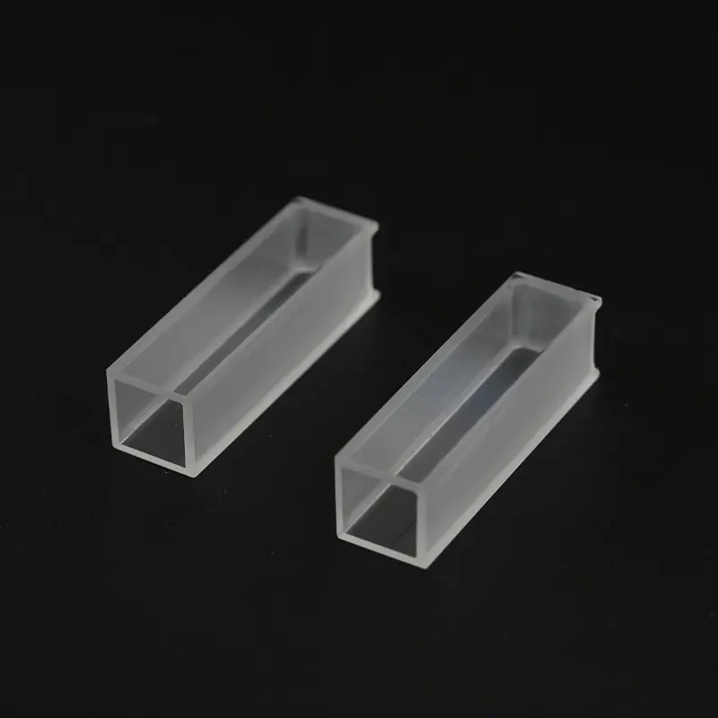
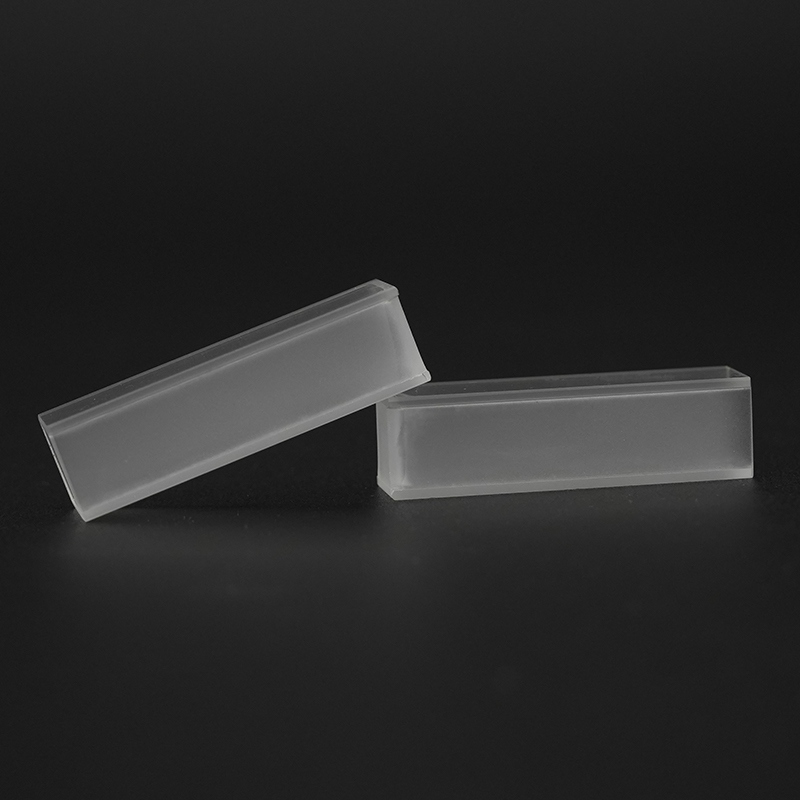 คิวเวตต์ซิลิกาควอตซ์ผสมสำหรับ UV-Vis และฟลูออเรสเซนต์ คูเวตต์ควอตซ์ที่หลอมรวมอย่างสมบูรณ์นี้เหมาะกับการดูดกลืนแสงและแสงฟลูออเรสเซนต์ตั้งแต่ 170 ถึง 2700 นาโนเมตร ใบหน้าแบบออพติคัลที่ขัดเงาและโครงสร้างที่แข็งแกร่งช่วยทำความสะอาดอย่างล้ำลึกและการตรวจสอบรายวันที่เชื่อถือได้ ดูผลิตภัณฑ์ →
 บทสรุปที่สำคัญ: ลำดับการล้างห้านาทีทุกวันป้องกันปัญหาพื้นฐานของ CD มากกว่า 90 เปอร์เซ็นต์ และควรถือเป็นส่วนที่ไม่สามารถต่อรองได้ในทุกเซสชันการวัด
การทำความสะอาดอย่างล้ำลึกสำหรับฟิล์มที่ฝังแน่นและตะกอน
เมื่อคิวเวทท์ไม่ผ่านการตรวจสอบยืนยันหลังจากโปรโตคอลรายวัน การทำความสะอาดอย่างล้ำลึกยิ่งขึ้นก็สมเหตุสมผล หลักการสำคัญคือการเลือกรีเอเจนต์ที่จะโจมตีแหล่งสะสมโดยไม่โจมตีควอตซ์ ซิลิกาผสมมีความทนทานทางเคมี แต่ไม่ทำลาย: ละลายในกรดไฮโดรฟลูออริกและกัดกร่อนอย่างช้าๆ ในอัลคาไลเข้มข้นที่ร้อน สูตรทำความสะอาดล้ำลึกทุกสูตรด้านล่างนี้คำนึงถึงขีดจำกัดเหล่านั้น
ฟิล์มโปรตีนจะถูกกำจัดออกได้ดีที่สุดโดยการย่อยอาหารมากกว่าการใช้สารเคมีที่รุนแรง แนวทางมาตรฐานคือสารละลายเปปซินที่ 1 ถึง 2 มก./มล. ในกรดไฮโดรคลอริก 0.1 โมลาร์ บ่มที่อุณหภูมิ 37 องศาเซลเซียส เป็นเวลาหนึ่งถึงสองชั่วโมง เอนไซม์จะแบ่งโปรตีนที่ถูกดูดซับออกเป็นเปปไทด์ขนาดเล็กซึ่งล้างออกได้ง่าย หลังจากการฟักไข่ ให้ล้างคิวเวตต์ให้สะอาดด้วยน้ำบริสุทธิ์พิเศษ จากนั้นจึงใช้เมทานอล จากนั้นทำให้แห้งด้วยไนโตรเจน อีกทางเลือกหนึ่งที่มีประสิทธิภาพคือสารละลายโซเดียมโดเดซิลซัลเฟต 0.5 ถึง 1 เปอร์เซ็นต์ อุ่นที่อุณหภูมิ 40 ถึง 50 องศาเซลเซียส ซึ่งละลายโปรตีนและไขมันได้ ต้องล้างให้สะอาดยิ่งขึ้นเพราะสาร SDS เองก็ดูดซับรังสี UV ไว้ได้
ตะกรันเกลือและแร่ธาตุตอบสนองต่อกรดเจือจาง กรดไนตริกที่ 1 ถึง 2 M เป็นตัวเลือกตามปกติ เนื่องจากกรดจะละลายสารตกค้างอนินทรีย์ส่วนใหญ่และไม่ทิ้งคราบไว้เลยหลังจากล้าง แช่ไว้ประมาณ 15 ถึง 30 นาทีที่อุณหภูมิห้อง อย่ารวมการทำความสะอาดด้วยกรดเข้ากับคลื่นโซนิค เนื่องจากการสั่นสะเทือนจะโจมตีขอบพื้นดินและเร่งปฏิกิริยา ฟิล์มอินทรีย์ทั่วไปจะถูกกำจัดออกโดยใช้ลำดับตัวทำละลาย: เมทานอล จากนั้น อะซิโตน จากนั้น เอทานอล และน้ำ ลำดับนี้เริ่มต้นด้วยตัวทำละลายที่ผสมน้ำได้ ละลายฟิล์มที่ไม่ชอบน้ำ และปิดท้ายด้วยตัวทำละลายที่ผสมกับน้ำเพื่อไม่ให้เกิดการตกตะกอน การล้างด้วยน้ำครั้งสุดท้ายจะขจัดคราบตัวทำละลายอินทรีย์ทั้งหมด อะซิโตนเพียงอย่างเดียวไม่เหมาะเป็นขั้นตอนแรกบนคิวเวตต์เปียกที่ปนเปื้อนเกลือ เนื่องจากอะซิโตนจะตกตะกอนเกลือและสามารถตรึงโปรตีนไว้บนพื้นผิวได้
คิวเวตแบบถอดประกอบได้ต้องใช้เทคนิคที่แตกต่างออกไป ควรแยกหน้าต่างออก ทำความสะอาดทีละบานบน PTFE แบบเรียบหรือพื้นผิวเช็ดในห้องสะอาดด้วยทิชชู่ไร้ขุยที่ชุบตัวทำละลาย แล้วเช็ดให้แห้งด้วยกระแสลมที่สะอาด การขันตัวเรือนโลหะให้แน่นเกินไปหลังการทำความสะอาดถือเป็นข้อผิดพลาดทั่วไป มันบิดเบือนหน้าต่างและสร้างการสะท้อนกลับของความเครียด ซึ่งเป็นสิ่งที่การตรวจวัดด้วยซีดีไม่สามารถทนได้ ประกอบกลับเข้าที่โดยขันให้แน่นบวกด้วยการหมุนเพียงเล็กน้อย และตรวจสอบความยาวเส้นทางด้วยวิธีการแก้ปัญหาอ้างอิง
| เงินฝาก | ขั้นตอนที่แนะนำ | เวลาติดต่อ | ไม่เคยใช้ |
|---|---|---|---|
| ฟิล์มโปรตีนที่ถูกดูดซับ | Pepsin 1-2 มก./มล. ใน 0.1 M HCl ที่ 37 องศาเซลเซียส | 1-2 ชม | ด่างเข้มข้นแบบร้อน |
| ระดับเกลือและแร่ธาตุ | กรดไนตริก 1-2 M ที่อุณหภูมิห้อง | 15-30 นาที | กรดไฮโดรฟลูออริกหรือน้ำยาทำความสะอาดฟลูออไรด์ |
| ฟิล์มลิพิด | สารลดแรงตึงผิวที่เป็นกลาง 1-2% ที่อุณหภูมิ 40 องศาเซลเซียส | 10-15 นาที | แผ่นขัดหรือผงขัด |
| ฝากอินทรีย์ทั่วไป | เมธานอล แล้วก็อะซิโตน แล้วก็เอทานอล แล้วก็น้ำ | 2-3 เล่ม of each | เช็ดให้แห้งด้วยกระดาษชำระ |
| ไม่ทราบหรือเงินฝากผสม | กรดไนตริก 10% ตามด้วยสารลดแรงตึงผิว 1-2% | ครั้งละ 30 นาที | ส่วนผสมที่ทำความร้อนได้เอง เช่น สารละลายปิรันย่าร้อน |
การทำความสะอาดอัลตราโซนิกสามารถเร่งทุกขั้นตอนข้างต้นได้ แต่สำหรับคิวเวตต์ชิ้นเดียวที่หลอมละลายอย่างสมบูรณ์เท่านั้น หากคิวเวทท์มีหน้าต่างที่ยึดด้วยซีเมนต์ ข้อต่ออาจรั่วหรือล้มเหลวได้ภายใต้การสั่นสะเทือนแบบอัลตราโซนิก เมื่อมีข้อสงสัย ให้ตรวจสอบเอกสารประกอบของซัพพลายเออร์ก่อนใส่คิวเวตต์ลงในอ่างอัลตราโซนิก คิวเวตต์ที่ทำจากควอตซ์ที่มีผนังหนาเทอะทะสามารถทนต่อรอบ sonication ในห้องปฏิบัติการมาตรฐานได้ 5 ถึง 10 นาที แต่เซลล์เรืองแสงที่มีผนังบางจะเปราะบางกว่า
บทสรุปที่สำคัญ: การทำความสะอาดแบบล้ำลึกเป็นการดำเนินการช่วยเหลือที่ปลอดภัยเฉพาะเมื่อรีเอเจนต์โจมตีสิ่งสะสม แต่ไม่ใช่โครงข่ายซิลิกา หากมีข้อสงสัย ให้เลือกสภาพที่อ่อนโยนกว่า
สิ่งที่ควรหลีกเลี่ยง: ข้อผิดพลาดในการทำความสะอาดที่สร้างความเสียหายให้กับคิวเวตควอตซ์
ความเสียหายจากคิวเวตต์ถาวรส่วนใหญ่ไม่ได้เกิดจากตัวอย่าง แต่เกิดจากความผิดพลาดในการทำความสะอาด ตัววัสดุเองมีความเสถียรทางเคมี แต่หน้าต่างขัดเงาบางๆ เสี่ยงต่อสารรีเอเจนต์ สารกัดกร่อน และการจัดการอย่างไม่ระมัดระวัง การทำความเข้าใจโหมดความล้มเหลวจะปกป้องทั้งข้อมูลคิวเวตต์และข้อมูลซีดี ตารางด้านล่างสรุปข้อผิดพลาดที่พบบ่อยที่สุดและผลที่ตามมา
| ความผิดพลาด | มีผลทันที | ความเสียหายระยะยาว |
|---|---|---|
| ทำความสะอาดด้วยกรดไฮโดรฟลูออริกหรือผลิตภัณฑ์ฟลูออไรด์ | หน้าต่างมีเมฆมากภายในไม่กี่นาที | ซิลิกาละลาย เซลล์จะต้องถูกทิ้ง |
| NaOH หรือ KOH เข้มข้นแบบร้อน | พื้นผิวเริ่มมีน้ำค้างแข็ง | การกัดแบบถาวรและการกระเจิงของแสงที่รุนแรง |
| แผ่นขัด ผงขัด หรือเครื่องมือโลหะ | มีรอยขีดข่วนเล็ก ๆ ปรากฏบนใบหน้า | สิ่งประดิษฐ์ซีดีที่กระจัดกระจายและชัดเจนซึ่งเลียนแบบวงดนตรีจริง |
| เช็ดให้แห้งด้วยกระดาษชำระ | มองเห็นการซ้อมและผ้าสำลี | รอยขีดข่วนแบบถาวรของควอตซ์ขัดเงา |
| อาบน้ำอัลตราโซนิกสำหรับเซลล์ซีเมนต์ | ข้อต่ออาจรั่วหรือเปิด | เซลล์สูญเสียการสอบเทียบความยาวเส้นทาง |
| ตัวทำละลายอินทรีย์ก่อนขจัดเกลือที่เป็นน้ำ | ผลึกเกลือตกตะกอน | คริสตัลสามารถขีดข่วนเซลล์ได้เมื่อเช็ด |
| ตัวอย่างที่ถูกทิ้งไว้ในเซลล์ข้ามคืน | เกิดเป็นฟิล์มโปรตีนแข็งและเกลือ | จำเป็นต้องทำความสะอาดอย่างรุนแรงซึ่งอาจทำให้พื้นผิวเสียหายได้ |
| ล้างด้วยน้ำประปา | ไอออนของโลหะสะสมบนซิลิกา | การดูดซับรังสียูวีและการชดเชยพื้นฐานเพิ่มเติม |
| การนึ่งเซลล์ที่ประกอบเข้าด้วยกันซ้ำแล้วซ้ำอีก | ความเครียดสะสมอย่างค่อยเป็นค่อยไป | การรีฟริงเจนต์ของความเครียดและเส้นฐานซีดีที่บิดเบี้ยว |
สารละลายปิรันย่าซึ่งเป็นส่วนผสมของกรดซัลฟิวริกเข้มข้นและไฮโดรเจนเปอร์ออกไซด์ บางครั้งใช้ในการทำความสะอาดไมโครอิเล็กทรอนิกส์ โดยทั่วไปไม่แนะนำให้ใช้คิวเวตต์แบบออปติคัล ปฏิกิริยาเป็นแบบคายความร้อนและสามารถกัดหรือเกิดเมฆควอทซ์ได้หากอุณหภูมิสูงขึ้น แต่ยังก่อให้เกิดอันตรายต่อความปลอดภัยอย่างร้ายแรงในห้องปฏิบัติการอีกด้วย หากคราบสกปรกต้านทานกรดไนตริกและผงซักฟอก คิวเวตต์จะถูกเปลี่ยนในราคาประหยัดมากกว่าเสี่ยงในอ่างปิรันย่า
บทสรุปที่สำคัญ: กฎทองของการดูแลคิวเวตต์นั้นเรียบง่าย: อย่าทำกับคิวเวตต์ในสิ่งที่คุณจะไม่ทำกับกล้องจุลทรรศน์
วิธีตรวจสอบว่าคิวเวทท์สะอาดอย่างแท้จริงก่อนทำการวัดค่าซีดี
การยืนยันเป็นขั้นตอนที่ห้องปฏิบัติการส่วนใหญ่ข้ามไป และเป็นขั้นตอนที่ตรวจพบปัญหาก่อนที่จะรวบรวมข้อมูล การตรวจสอบด้วยภาพเพียงอย่างเดียวไม่เพียงพอ เนื่องจากภาพยนตร์ที่เกี่ยวข้องกับซีดีส่วนใหญ่เป็นภาพยนตร์ที่มีชั้นเดียว ลำดับต่อไปนี้จะเปลี่ยนจากการตรวจสอบอย่างง่ายไปสู่การทดสอบโดยใช้เครื่องมือ
- การตรวจสอบด้วยสายตา: ถือคิวเวตต์เปล่าที่สะอาดในมุมหนึ่งกับพื้นหลังที่สว่าง และมองหาเส้นริ้ว คริสตัล เส้นใย หรือความขุ่นบนผิวหน้าเลนส์
- การทดสอบฟิล์มน้ำ: เติมน้ำบริสุทธิ์พิเศษแล้วเทออก พื้นผิวซิลิกาหลอมละลายที่สะอาดจะยังคงเปียกอย่างสมบูรณ์ ในขณะที่ฟิล์มที่ไม่ชอบน้ำจะทำให้น้ำกลายเป็นเม็ดบีด
- การสแกนการส่งผ่านรังสียูวี: เติมน้ำบริสุทธิ์พิเศษและสแกนจากอากาศที่ความสูง 190 ถึง 400 นาโนเมตร ค่าการดูดกลืนแสงปรากฏที่ 220 นาโนเมตรควรต่ำกว่า 0.05 AU และค่าที่ 280 นาโนเมตรต่ำกว่า 0.01 AU
- การทดสอบพื้นฐานจาก CD: วางคิวเวตต์ที่เติมน้ำไว้ในเครื่องมือ และบันทึกเส้นพื้นฐานเหนือช่วงการทำงาน เส้นพื้นฐานควรคงที่ภายในประมาณบวกหรือลบ 0.5 mdeg และแรงดันไดโนดควรคงอยู่ที่ค่าปกติสำหรับรุ่นอุปกรณ์
- ทำซ้ำการซ้อนทับ: รวบรวมการสแกนตัวอย่างเดียวกันสามครั้งติดต่อกัน ควรวางซ้อนภายในระดับเสียง และการเปลี่ยนแปลงหรือการเลื่อนใด ๆ บ่งชี้ว่ามีฟิล์มเคลื่อนไหวหรือฟองอากาศ
- การตรวจสอบมาตรฐานอ้างอิง: สำหรับเซลล์ใหม่หรือหลังจากประกอบเซลล์แบบถอดประกอบกลับคืนได้ ให้ตรวจสอบด้วยข้อมูลอ้างอิงที่ได้รับการรับรอง เช่น กรดการบูรซัลโฟนิก และยืนยันว่าแถบความถี่ใกล้ 290.5 นาโนเมตรตรงกับค่าที่คาดหวังสำหรับความยาวเส้นทาง
คิวเวตต์ยังคงสามารถบันทึกได้
| ควรเปลี่ยนคิวเวตต์
|
ค่าเกณฑ์ข้างต้นเป็นค่าเริ่มต้นในทางปฏิบัติที่ใช้ในห้องปฏิบัติการโปรตีนซีดี คิวเวตต์ที่ผ่านการตรวจสอบทั้งหกครั้งจะไม่ใช่ปัจจัยจำกัดในการวัดของคุณ หากการตรวจสอบล้มเหลว ให้ทำความสะอาดแบบล้ำลึกอีกครั้ง หากล้มเหลวอีกครั้ง ให้เปลี่ยนเซลล์แทนที่จะใช้เวลาหลายชั่วโมงกับสาเหตุที่สูญเสียไป
บทสรุปที่สำคัญ: การตรวจสอบยืนยันสองนาทีต่อคิวเวตต์ช่วยปกป้องชั่วโมงการวัดและป้องกันไม่ให้ชุดข้อมูลทั้งหมดสูญเปล่า
การเลือกคิวเวตต์ควอตซ์ที่เหมาะสมเพื่อลดภาระในการทำความสะอาดและสิ่งแปลกปลอมทางแสง
แนวทางการทำความสะอาดที่ดีที่สุดเริ่มต้นจากการตัดสินใจซื้อ คิวเวตต์ที่ระบุอย่างถูกต้องสำหรับซีดีจะทำความสะอาดได้เร็วขึ้น ตรวจสอบได้ง่ายขึ้น และสร้างเส้นฐานที่ทำซ้ำได้มากขึ้นตลอดอายุการใช้งาน การตัดสินใจสามประการมีอิทธิพลเหนือ: เกรดวัสดุ การสร้างเซลล์ และความยาวเส้นทาง
เกรดวัสดุจะกำหนดความยาวคลื่นที่สั้นที่สุดที่ใช้งานได้ สำหรับฟาร์-ยูวีซีดีที่ต่ำกว่า 210 นาโนเมตร ให้ระบุซิลิกาผสมสังเคราะห์ JGS1 พร้อมการส่งผ่านเอกสารในช่วง 170 ถึง 220 นาโนเมตร ควอตซ์ผสม JGS2 เป็นที่ยอมรับสำหรับการวัดที่อยู่เหนือ 210 นาโนเมตร ควรระบุเกรดไว้ในใบรับรองของซัพพลายเออร์ หากไม่เป็นเช่นนั้น แสดงว่าคิวเวตต์อาจไม่ได้มีไว้สำหรับซีดี โครงสร้างเป็นตัวกำหนดความเข้ากันได้ในการทำความสะอาด เซลล์ชิ้นเดียวที่หลอมรวมอย่างสมบูรณ์ไม่มีกาวอินทรีย์ และสามารถแช่ในน้ำยาทำความสะอาดใดๆ ที่ระบุไว้ข้างต้นได้ รวมถึงคลื่นเสียงสั้นๆ ด้วย เซลล์ซีเมนต์มีราคาถูกกว่า แต่ไม่เหมาะสำหรับการทำความสะอาดแบบเข้มข้น เซลล์แบบถอดประกอบได้ช่วยให้เข้าถึงหน้าต่างได้ดีที่สุดเพื่อการทำความสะอาดอย่างล้ำลึก และเป็นตัวเลือกมาตรฐานสำหรับความยาวเส้นทางที่สั้นมาก การ์ดด้านล่างสรุปความยาวเส้นทางมาตรฐานและแอปพลิเคชันซีดีที่แนะนำ
ทางเดิน 0.1 มม
ใช้สำหรับตัวอย่างโปรตีนเข้มข้นและตรวจวัดได้ถึง 185 นาโนเมตร ปริมาตรตัวอย่างประมาณ 10-20 ไมโครลิตร มักจำหน่ายเป็นเซลล์แบบถอดประกอบได้เพื่อหลีกเลี่ยงการติดฟอง
ทางเดิน 0.5 มม
การประนีประนอมที่ดีสำหรับ Far-UV CD ระหว่าง 185 ถึง 220 นาโนเมตร ปริมาตรประมาณ 50-100 µL. แนะนำสำหรับสารละลายโปรตีน 5 ถึง 20 มก./มล.
ทางเดิน 1 มม
ความยาวเส้นทางซีดีที่พบบ่อยที่สุด ปริมาตรประมาณ 100-200 µL. เหมาะสำหรับการตรวจวัดแบบ Far-UV และ Near-UV ด้วยความเข้มข้นของตัวอย่างในระดับปานกลาง
ทางเดิน 2 มม
ขยายความไวของแผ่นซีดีใกล้รังสียูวีในขณะที่ยังคงสามารถทำงานได้กับรังสียูวีไกล ปริมาณประมาณ 200-400 µL.
เส้นทาง 5 มม
ดีที่สุดสำหรับซีดีใกล้ UV ของอะโรมาติกตกค้างและโคแฟคเตอร์ที่สูงกว่า 250 นาโนเมตร ปริมาณประมาณ 500-1,000 µL.
ทางเดิน 10 มม
ความยาวเส้นทางมาตรฐานสำหรับ UV-Vis และ CD ใกล้ UV โดยทั่วไปจะดูดซับรังสี UV ต่ำกว่า 200 นาโนเมตรด้วยบัฟเฟอร์ที่เป็นน้ำมากเกินไป ปริมาณประมาณ 1-2 มล.
นอกเหนือจากความยาวเส้นทางแล้ว ให้พิจารณาเส้นทางแสงที่สมบูรณ์ ผิวหน้าของหน้าต่างจะต้องได้รับการขัดเงาให้มีข้อกำหนดการเจาะรอยขีดข่วนต่ำ และคิวเวตต์ต้องปราศจากการรีฟริงก์ที่เกิดจากความเครียด การทดสอบง่ายๆ ระหว่างโพลาไรเซอร์แบบไขว้เผยให้เห็นความเครียดที่มากเกินไป คิวเวตที่มีผนังสีดำหรือหน้าต่างปิดบังมีประโยชน์สำหรับการศึกษาการเรืองแสง แต่ไม่จำเป็นสำหรับซีดีมาตรฐาน โครงสร้างที่ปราศจากโลหะถือเป็นข้อได้เปรียบหากจะใช้คิวเวตต์กับบัฟเฟอร์ที่โจมตีโครงโลหะ
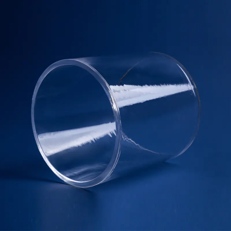
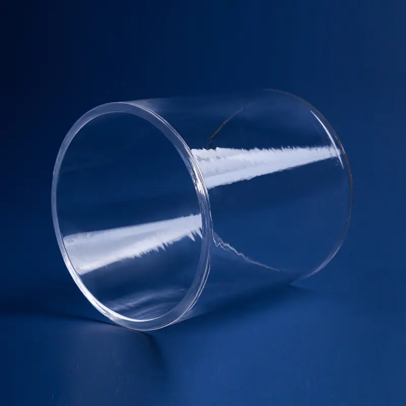
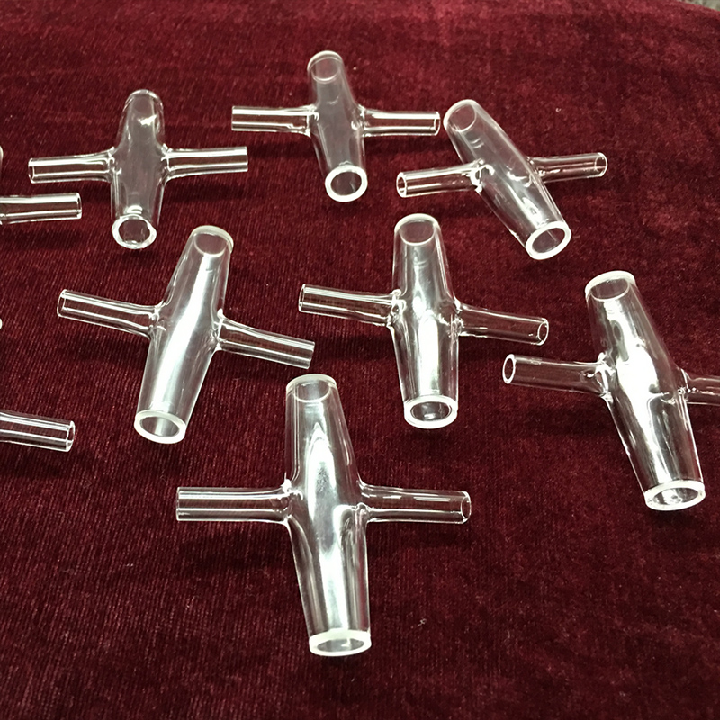 คิวเวตแก้วควอตซ์ใสสำหรับเครื่องมือวัดแสง ออกแบบมาสำหรับเครื่องสเปกโตรโฟโตมิเตอร์ UV-Vis และฟลูออเรสเซนซ์ คิวเวตต์แบบควอตซ์นี้ทนทานต่อสารเคมีส่วนใหญ่และทนทานต่ออุณหภูมิการทำงานที่สูง ความชัดเจนและความทนทานของแสงช่วยให้ถือและจัดเก็บในระยะยาวได้จริง ดูผลิตภัณฑ์ →
 บทสรุปที่สำคัญ: คิวเวตต์ที่ระบุอย่างถูกต้องเป็นการอัพเกรดที่ถูกที่สุดในห้องปฏิบัติการซีดี โดยช่วยลดเวลาในการทำความสะอาด ทำให้ค่าพื้นฐานคงที่ และยืดอายุการใช้งานของเซลล์หลายปี
การจัดเก็บ การจัดการ และการบำรุงรักษาตามปกติเพื่ออายุการใช้งานคิวเวตต์ที่ยาวนาน
วิธีจัดเก็บและจัดการคิวเวตต์ระหว่างการวัดต่างๆ จะเป็นตัวกำหนดความถี่ที่ต้องทำความสะอาด หน้าต่างซิลิกาที่หลอมละลายจะดึงฟิล์มจากฝุ่นในอากาศ ลายนิ้วมือ และแม้กระทั่งไอระเหยของตัวทำละลายอินทรีย์ที่จัดเก็บไว้ในตู้เดียวกัน พฤติกรรมต่อไปนี้จะทำให้คิวเวตต์คงสภาพการมองเห็นได้นานหลายปี
กฎการจัดการ: จับคิวเวตไว้ที่ผนังด้านข้างที่มีน้ำค้างแข็งหรือขอบด้านบนเท่านั้น สวมถุงมือไนไตรล์ชนิดไม่มีแป้ง และใช้แหนบปลายแหลม PTFE เมื่อเคลื่อนย้ายเซลล์ อย่าสัมผัสใบหน้าออพติคอลที่ขัดเงา ใช้กล่องคิวเวทท์แบบบุนุ่มโดยเฉพาะสำหรับการจัดเก็บและขนส่ง และเก็บเซลล์ให้ตั้งตรงหรือวางราบบนพื้นผิวที่สะอาดไม่เป็นขุย หน้าสัมผัสระหว่างกระจกกับกระจก ซึ่งเกิดขึ้นเมื่อคิวเวตต์วางซ้อนกันในลิ้นชัก เป็นสาเหตุหนึ่งที่มักทำให้เกิดเศษที่ขอบ
กฎการจัดเก็บ: เก็บคิวเวตให้ว่างและแห้งโดยถอดฝาหรือตัวหยุดออกหรือวางหลวมๆ เพื่อไม่ให้ความชื้นควบแน่นภายใน อย่าเก็บคิวเวตที่แช่อยู่ในตัวทำละลาย เมทานอล หรือน้ำ การสัมผัสตัวทำละลายเป็นเวลานานจะทำให้เซลล์ซีเมนต์นิ่มลง และน้ำนิ่งจะกระตุ้นให้เกิดฟิล์มสาหร่ายหรือแบคทีเรีย ทำความสะอาดคิวเวตต์ที่เก็บไว้อีกครั้งหลังจากไม่ได้ใช้งานเป็นเวลานานกว่าหนึ่งสัปดาห์ เนื่องจากแม้แต่การจัดเก็บแบบไร้ฝุ่นก็ช่วยให้ฟิล์มอินทรีย์บางๆ สะสมบนซิลิกาที่หลอมละลายได้
ตารางการบำรุงรักษา: ตรวจสอบคิวเวตต์ซีดีทั้งหมดทุกสัปดาห์เพื่อดูฟิล์มและรอยขีดข่วนที่มองเห็นได้ ทำความสะอาดอย่างล้ำลึกเป็นรอบทุกเดือน หรือเร็วกว่านั้นหากการตรวจสอบพื้นฐานไม่สะอาดอีกต่อไป เปลี่ยนคิวเวตต์หลังจากการตรวจสอบล้มเหลวติดต่อกันสองครั้ง หากห้องปฏิบัติการใช้งานซีดีทุกวัน ให้เก็บคิวเวตที่ตรงกันอย่างน้อยสองคิวเวตต่อระบบบัฟเฟอร์ เพื่อให้สามารถทำความสะอาดคิวเวตหนึ่งได้ในขณะที่อีกคิวใช้งานอยู่ การสแกนการส่งผ่าน UV-Vis อย่างรวดเร็วบนคิวเวตต์เปล่าที่สะอาดทุกๆ สองสามเดือนจะบันทึกว่าหน้าต่างมีอายุอย่างช้าๆ หรือไม่
บทสรุปที่สำคัญ: จัดการคิวเวตเป็นส่วนประกอบเชิงแสงที่มีความแม่นยำ จัดเก็บเหมือนเครื่องมือที่เปราะบาง และทำความสะอาดอย่างล้ำลึกตามกำหนดเวลา แทนที่จะรอให้เส้นฐานที่ปนเปื้อนปรากฏขึ้น
การจัดหาคิวเวตควอตซ์จากผู้ผลิตหรือซัพพลายเออร์ที่เชื่อถือได้
สำหรับห้องปฏิบัติการ ผู้จัดจำหน่าย และผู้ซื้อ OEM ต้นทุนระยะยาวของคิวเวตต์จะขึ้นอยู่กับความสม่ำเสมอและความสามารถในการทำความสะอาด ไม่ใช่ราคาเริ่มต้น คิวเวตต์จากผู้ผลิตควอตซ์ที่มีวัตถุดิบเกรดออพติคัลที่ได้รับการควบคุมและการประมวลผลที่จัดทำเป็นเอกสารจะทำความสะอาดซ้ำได้เป็นเวลาหลายปี เซลล์ราคาถูกที่มีเกรดไม่แน่นอนจะทำให้เกิดการเปลี่ยนแปลงพื้นฐานที่ละเอียดอ่อนซึ่งยากต่อการติดตาม นี่คือเหตุผลที่การจัดซื้อจัดจ้างควรได้รับการปฏิบัติเป็นส่วนหนึ่งของระบบคุณภาพ
เมื่อคุณขอใบเสนอราคาจากผู้ผลิตหรือซัพพลายเออร์คิวเวตต์ควอตซ์ ให้ขอเอกสารเฉพาะ เกรดวัสดุควรระบุเป็น JGS1, JGS2 หรือการกำหนดสากลที่เทียบเท่า ซัพพลายเออร์ควรจัดเตรียมกราฟการส่งผ่านที่วัดได้ในช่วง 190 ถึง 260 นาโนเมตร ค่าเผื่อความยาวเส้นทางที่บวกหรือลบ 0.01 มม. หรือดีกว่า และยืนยันว่าหน้าต่างปราศจากความเครียด สำหรับงานซีดี การรับรองแบบไร้ความเครียดมีความสำคัญมากกว่า UV-Vis เนื่องจากการหักเหของแสงจะสร้างสิ่งแปลกปลอมทางแสงโดยตรง ขอความเข้ากันได้ในการทำความสะอาดของเซลล์ด้วย: ไม่ว่าจะหลอมรวมหรือซีเมนต์อย่างสมบูรณ์ และไม่ว่าจะมีโซนิคหรือไม่
สำหรับผู้ค้าส่งและผู้จัดจำหน่ายที่ซื้อในปริมาณมาก บรรจุภัณฑ์และการตรวจสอบย้อนกลับมีความสำคัญพอๆ กับออพติก เซลล์ควรมาถึงในกล่องป้องกันแต่ละอันพร้อมหมายเลขล็อตบนฉลาก ความสอดคล้องกันแบบแบตช์ต่อแบตช์เป็นสิ่งสำคัญ ใบรับรองสำหรับแต่ละแบทช์จะป้องกันสถานการณ์ที่คิวเวตหนึ่งมีพฤติกรรมแตกต่างจากที่อื่น ควรถามว่าผู้ผลิตเสนอขนาดที่กำหนดเอง การออกแบบแบบถอดประกอบได้ และหน้าต่างที่กำหนดเองโดยไม่มีปริมาณการสั่งซื้อขั้นต่ำจำนวนมากหรือไม่ Yancheng Mingyang Quartz Products Co., Ltd. เป็นผู้ผลิตผลิตภัณฑ์ควอตซ์ใน Yancheng ประเทศจีน และทำหน้าที่เป็นโรงงานผลิตของ Jinzhou Mingde Quartz Glass Co., Ltd.; บริษัทผลิตคิวเวตต์ควอตซ์ เครื่องควอตซ์ในห้องปฏิบัติการ แผ่นควอตซ์แบบออปติคัล และส่วนประกอบควอตซ์สั่งทำสำหรับลูกค้าห้องปฏิบัติการและลูกค้าอุตสาหกรรมทั่วโลก ความสัมพันธ์กับผู้ผลิตโดยตรงช่วยให้ผู้ซื้อสามารถควบคุมเกรด การรับรอง และการผลิตแบบกำหนดเองได้
บทสรุปที่สำคัญ: ซื้อคิวเวตสำหรับซีดีพร้อมใบรับรอง ไม่ใช่แค่หมายเลขแค็ตตาล็อก การยืนยันเกรดและแบบไร้ความเครียดมีค่าใช้จ่ายเพียงเล็กน้อยในการซื้อ แต่ช่วยประหยัดการแก้ไขปัญหาจำนวนมากในภายหลัง
คำถามที่พบบ่อยเกี่ยวกับการทำความสะอาดคิวเวตต์ควอตซ์สำหรับ CD Spectroscopy
มีวิธีที่ดีที่สุดในการทำความสะอาดคิวเวตต์ควอตซ์สำหรับ CD สเปกโทรสโกปีหรือไม่วิธีการที่เชื่อถือได้มากที่สุดคือลำดับมาตรฐาน: ระบายตัวอย่าง, ล้างด้วยบัฟเฟอร์, ล้างด้วยน้ำบริสุทธิ์พิเศษ, ล้างด้วยผงซักฟอกที่เข้ากันได้กับควอทซ์ที่เป็นกลาง 1-2 เปอร์เซ็นต์, ล้างอีกครั้งด้วยน้ำ, ล้างด้วยเมทานอลหรือเอทานอล และเช็ดให้แห้งด้วยไนโตรเจน จุดสำคัญคืออย่าปล่อยให้ตัวอย่างแห้งในเซลล์ และต้องตรวจสอบผลลัพธ์ด้วยน้ำพื้นฐาน 220 นาโนเมตร |
ฉันสามารถใช้อ่างอัลตราโซนิกเพื่อทำความสะอาดคิวเวตต์ซีดีควอตซ์ได้หรือไม่เฉพาะในกรณีที่คิวเวตต์มีโครงสร้างชิ้นเดียวที่หลอมรวมอย่างสมบูรณ์เท่านั้น คิวเวตต์แบบซีเมนต์หรือแบบสองส่วนสามารถแยกหรือรั่วไหลได้ภายใต้การสั่นสะเทือนแบบอัลตราโซนิก ตรวจสอบเอกสารของผู้ผลิต หากมีข้อสงสัย ให้ใช้การแช่ด้วยตนเองแทน |
ฉันจะเอาฟิล์มโปรตีนที่ไม่หลุดออกพร้อมกับผงซักฟอกได้อย่างไรใช้การย่อยด้วยเอนไซม์ เช่น เปปซิน ที่ 1 ถึง 2 มก./มล. ในกรดไฮโดรคลอริก 0.1 โมลาร์ ที่อุณหภูมิ 37 องศาเซลเซียส เป็นเวลาหนึ่งถึงสองชั่วโมง ล้างออกให้สะอาดหลังจากนั้น หลีกเลี่ยงโซเดียมไฮดรอกไซด์ร้อนซึ่งจะกัดกร่อนหน้าต่างควอตซ์ |
ลายนิ้วมือส่งผลต่อการวัดค่าซีดีจริงหรือ?ใช่และน่าทึ่งมาก สารตกค้างจากลายนิ้วมือประกอบด้วยกรดอะมิโนและกรดไขมันที่ดูดซับได้ต่ำกว่า 250 นาโนเมตร การสัมผัสโดยไม่ได้ตั้งใจเพียงครั้งเดียวสามารถเพิ่ม 0.05 ถึง 0.1 AU ที่ 220 นาโนเมตร และทำลายความเรียบของเส้นพื้นฐาน หยิบคิวเวตต์ชิดผนังฝ้าเสมอขณะสวมถุงมือชนิดไม่มีแป้ง |
ควรทำความสะอาดคิวเวตต์ควอทซ์สำหรับ CD spectroscopy บ่อยแค่ไหนหลังการใช้งานทุกครั้ง โดยเฉพาะอย่างยิ่งเมื่อตัวอย่างมีโปรตีน ลิพิด หรือเกลือ ระบายและล้างคิวเวตต์ทันทีหลังการตรวจวัด คิวเวตที่ไม่ได้ใช้เป็นเวลานานกว่าหนึ่งสัปดาห์ควรทำความสะอาดอีกครั้งก่อนตั้งค่าการตรวจวัดครั้งถัดไป |
เมื่อใดจึงควรเปลี่ยนคิวเวตต์ควอตซ์แทนการทำความสะอาดเปลี่ยนคิวเวตต์เมื่อการทำความสะอาดแบบล้ำลึกสองครั้งติดต่อกันไม่คืนสภาพพื้นฐาน เมื่อมีรอยขีดข่วนที่มองเห็นได้ข้ามพื้นผิวที่มองเห็นได้ เมื่อเซลล์รั่ว หรือเมื่อคราบบนพื้นผิวที่แข็งตัวบ่งบอกถึงการกัดกร่อน ซิลิกาคิวเวทต์หลอมที่ได้รับการจัดการอย่างถูกต้องสามารถมีอายุการใช้งานหลายปี สาเหตุปกติของความล้มเหลวตั้งแต่เนิ่นๆ ได้แก่ กรดไฮโดรฟลูออริก อัลคาไลเข้มข้นที่ร้อน และการเสียดสีทางกล |
บทสรุปที่สำคัญ: กลยุทธ์การทำความสะอาดที่ดีที่สุดคือการป้องกัน: ล้างทันที ตรวจสอบทุกวัน และเปลี่ยนเฉพาะเมื่อการทำความสะอาดไม่สามารถคืนสภาพพื้นผิวเลนส์ได้
แหล่งข้อมูลที่เกี่ยวข้องและข้อมูลผลิตภัณฑ์
สำหรับข้อมูลจำเพาะของคิวเวทท์ รูปทรงมาตรฐาน และเกรดควอตซ์ที่มีในกลุ่มผลิตภัณฑ์ โปรดดูที่ คิวเวตต์ควอตซ์สำหรับสเปกโทรสโกปี หน้า
หากแอปพลิเคชันซีดีของคุณต้องการความยาวเส้นทางที่ไม่เป็นมาตรฐาน การออกแบบแบบถอดประกอบได้ หรือเกรดซิลิกาผสมเฉพาะ การผลิตเครื่องควอทซ์แบบกำหนดเอง บริการรองรับการผลิตแบบครั้งเดียวและแบบเป็นชุด
ในฐานะผู้ผลิตและจำหน่ายผลิตภัณฑ์ควอตซ์ Yancheng Mingyang Quartz Products Co., Ltd. ผลิตคิวเวตต์ควอตซ์ เครื่องควอตซ์ในห้องปฏิบัติการ แผ่นควอตซ์แบบออปติคอล และส่วนประกอบที่เกี่ยวข้องสำหรับสเปกโทรสโกปีซีดี การวิเคราะห์ UV-Vis และการใช้งานด้านแสงอื่นๆ บริษัทดำเนินงานในฐานะโรงงานผลิตของ Jinzhou Mingde Quartz Glass Co., Ltd. โดยให้บริการลูกค้าในห้องปฏิบัติการ อุตสาหกรรม และลูกค้าขายส่งทั่วตลาดทั่วโลก
สินค้าแนะนำ
ลิขสิทธิ์ © Yancheng Mingyang ควอตซ์โปรดักส์ จำกัด สงวนลิขสิทธิ์.
ผู้ผลิตขายส่งผลิตภัณฑ์ควอตซ์ โรงงานแก้วควอตซ์